ColabMed: Automated TICI Scoring with DL
Automated TICI Scoring with DL
![]()
This repository is a fork of https://github.com/IPMI-ICNS-UKE/DeepTICI.
I have streamlined the source code, enabling it for the prediction of Thrombolysis in Cerebral Infarction (TICI) scores for a given Digital Subtraction Angiography (DSA) series with two views (i.e., lateral and frontal), accounting for the original M1 occlusion.

Requirements
All you need is a Google account with access to Colaboratory.
Installation
No coding expertise is required, and there’s no need for manual software installation on your part.
Usage
Simply follow the video demonstration to input the DSA sequence and obtain your TICI score. The only consideration is to ensure that the ‘ImageType’ in the Dicom file properties you enter contains the keywords ‘BIPLANE A’ and ‘BIPLANE B’ for clear differentiation between lateral and frontal views.
Label Mapping
1
2
3
4
5
6
7
8
label_mapping = {
    0: 'TICI 0',
    1: 'TICI 0',
    2: 'TICI 2a',
    3: 'TICI 2b',
    4: 'TICI 3',
    5: 'TICI 3'
}
Modified Treatment in Cerebral Ischemia Scale
| mTICI | Definitions |
|---|---|
| Grade 0 | No perfusion |
| Grade 1 | Antegrade reperfusion past the initial occlusion, but limited distal branch filling with little or slow distal reperfusion |
| Grade 2a | Antegrade reperfusion of less than half of the occluded target artery previously ischemic territory (eg, in 1 major division of the MCA and its territory) |
| Grade 2b | Antegrade reperfusion of more than half of the previously occluded target artery ischemic territory (eg, in 2 major divisions of the MCA and their territories) |
| Grade 3 | Complete antegrade reperfusion of the previously occluded target artery ischemic territory, with the absence of visualized occlusion in all distal branches |
MCA indicates middle cerebral artery; and mTICI, Modified Treatment in Cerebral Ischemia Scale.
American Society of Interventional and Therapeutic Neuroradiology Collateral Grading System
| ACG Grades | Definitions |
|---|---|
| Grade 0 | No collaterals visible to the ischemic site |
| Grade 1 | Slow collaterals to the periphery of the ischemic site, with persistence of some of the defect |
| Grade 2 | Rapid collaterals to the periphery of the ischemic site, with persistence of the defect, and only to a portion of the ischemic territory |
| Grade 3 | Collaterals with slow but complete angiographic blood flow of the ischemic bed by the late venous phase |
| Grade 4 | Complete and rapid collateral blood flow to the vascular bed in the entire ischemic territory by retrograde perfusion |
| N/A | Not applicable based on the territory or injections available |
ACG indicates the American Society of Interventional and Therapeutic Neuroradiology collateral grading.
Discussion
This is the original paper along with the corresponding flowchart:
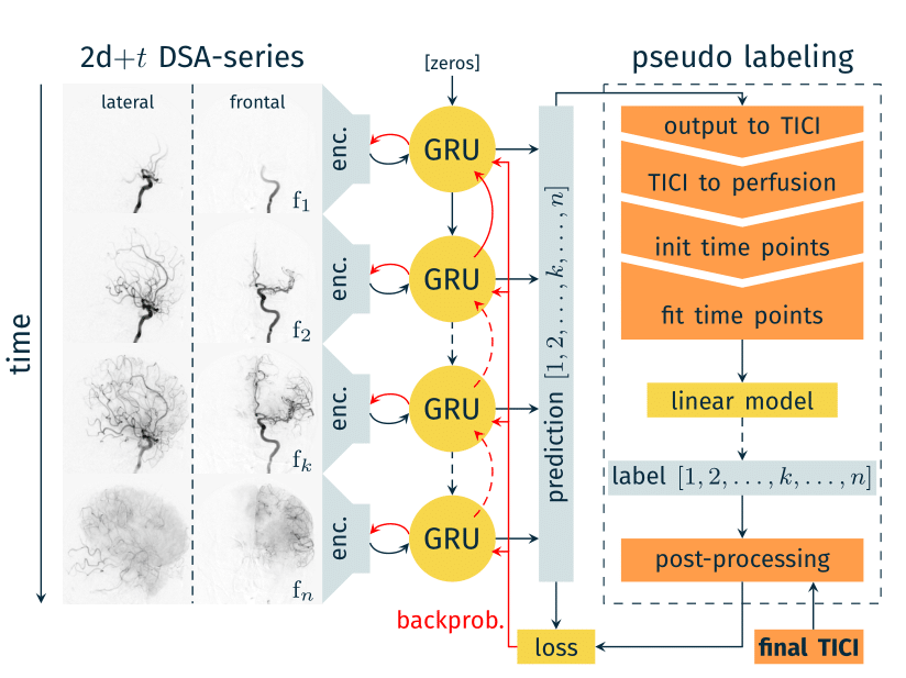
While adopting a time-based classification is indeed a sensible approach, I am inclined to believe that the contributions of f1, f2, fn in the plot may not exert a substantial impact on the scores, at least not to the extent observed with the contribution of fk.
For seasoned clinicians, do you find it plausible to derive a TICI score solely from fk (or including fk-1, fk, fk+1)? This simplification could hold profound consequences for AI models.
Your insights and contributions to the open-source code are highly valued. Please feel free to share your ideas.
Kindly refrain from utilizing it for commercial purposes.